MRI and Imaging Information
Brain imaging at WERC is done with magnetic resonance imaging (MRI) scanners, including the Connectome Skyra used in the landmark Human Connectome Project at Washington University, St. Louis. This is a state-of-the-art scanner, customized to allow exceptionally high structural detail of brain fiber tracts. MRI scanning allows us to assess the brains of individuals including those with schizophrenia and bipolar disorder and healthy participants. We use a variety of computational imaging methods, including those assessing brain:
-
Structural Connectivity:
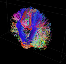
This is studied using diffusion MRI that gives insight into how regions of the brain are physically wired together into networks that communicate electrical signals through the white matter. Diffusion MRI is based upon measuring the diffusion of molecules, mainly water, through tissue. Diffusion tractography is a method for identifying anatomical connections in the living human brain. -
Resting-State Functional Connectivity:
Resting state functional MRI (R-fMRI) is a powerful method for evaluating regional interactions that occur when a subject is not performing an explicit task. Brain activity is present even in the absence of an externally prompted task, and this can be detected by measuring spontaneous fluctuations in the Blood Oxygenation Level-Dependent Signal (BOLD). R-fMRI can reveal a number of networks representing regions with common patterns of synchronous activity. -
Task-Related Functional Connectivity:
Another way to try to identify different subdivisions of the human brain is using task fMRI. This approach examines brain regions whose BOLD activity changes when people are asked to process different kinds of information (e.g. words, pictures, sounds), or use different types of thinking skills (e.g. memory, language generation). Results can help guide, validate and interpret the result of the connectivity analyses obtained using R-fMRI and diffusion MRI. -
Volume, Shape and Folding Patterns:
Using structural MRI methods, the size and shape of different brain regions (e.g. hippocampus, thalamus, basal ganglia) can be determined. We can also study the wrinkles or folding patterns of the cerebral cortex. This folding pattern varies from one person to the other. -
Other:
With advanced scanning and data-analysis, the myelin content of brain fibers can be studied. Using new MRI methods developed at Washington University, it may also be possible to quantify the degree of brain tissue degeneration and derive information about the potential causes of brain abnormalities.
Through our studies we hope to improve the classification of psychiatric disorders, which currently is based on clinical symptoms only. We are conducting diverse clinical assessments to better understand the relevance of brain imaging findings. These assessments include those measuring psychosis, depression, mania, cognition, social functioning, perceived stress, personality traits and disability.